


Meeting Reviews
CCAS 2017 Annual Meeting Review: Morning Sessions
By Rania K. Abbasi, MD
Riley Hospital for Children
Indianapolis, IN
2017 marks the 10th anniversary of the CCAS Annual Meeting. This year’s meeting took place in Austin, Texas, the Live Music Capital of the World. Program chair, Scott G. Walker, MD (Riley Hospital for Children, Indianapolis), and his co-chairs Luis Zabala, MD (UT-Southwestern/Children’s Medical Center, Dallas) and Kirsten Odegard, MD (Boston Children’s Hospital, Boston) organized a captivating series of speakers on a wide variety of topics, from genetics of CHD, congenitally corrected transposition of the great arteries, innovations in the cath lab, and updates on thoracic surgery. Several new features were debuted, including morning PBLD sessions, an “ask the speaker” feature on the mobile meeting guide, and a transesophageal echocardiography workshop featuring Vimedix simulators.
The morning began with the new PBLD session. The topics included point-of-care coagulation testing with Jumbo Williams, MB, ChB (Lucille Packard Children’s Hospital, Palo Alto) and Laura Downey, MD (Children’s Healthcare of Atlanta, Atlanta), the Norwood patient for noncardiac surgery with Susan Staudt, MD and Lindsey Loveland Baptist, MD (Children’s Hospital of Wisconsin, Milwaukee), anesthetic considerations for Williams syndrome with Susan Nicolson, MD and Kelly Grogan, MD (CHOP, Philadelphia), fontan conversion surgery with Emad Mossad, MD and Premal Trivedi, MD (Texas Children’s Hospital, Houston), and managing risk in the cath lab with Kirsten Odegard, MD and Viviane Nasr, MD (Boston Children’s Hospital, Boston). Nearly all groups were sold out!
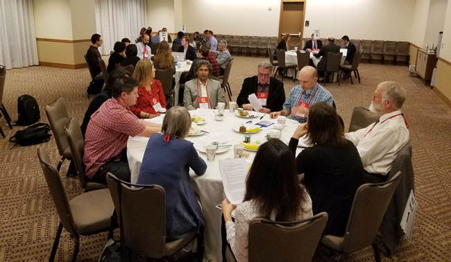
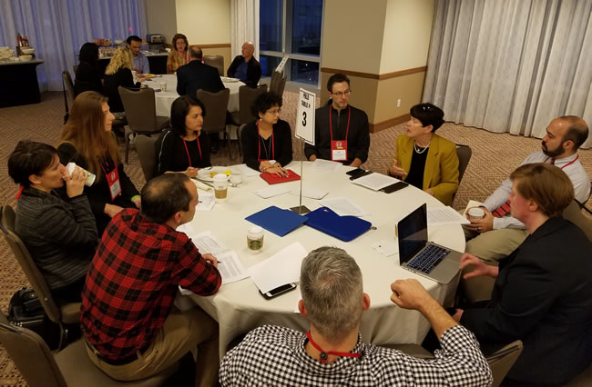
Morning PBLD sessions
The morning general session was kicked off by outgoing CCAS president, Emad B. Mossad, who thanked outgoing board of directors members Helen Holtby, MBBS, FRCP (The Hospital for Sick Children, Toronto), Kirsten Odegard, Courtney Hardy, MD (Lurie Children’s Hospital, Chicago), Scott Walker, and Jumbo Williams. Dr. Mossad then passed the torch to our new CCAS president, Nina Guzzetta (Children’s Healthcare of Atlanta, Atlanta), who introduced the continuing board members Jeremy Geiduscheck, MD (Seattle Children’s Hospital, Seattle) and Luis Zabala, MD (UT Southwestern Children’s Medical Center, Dallas) and welcomed incoming board members Susan Nicolson, Viviane Nasr (Boston Children’s Hospital, Boston), Greg Latham, MD (Seattle Children’s Hospital, Seattle), Jim Spaeth (Cincinnati Children’s Hospital, Cincinnati), and David Vener (Texas Children’s Hospital, Houston).
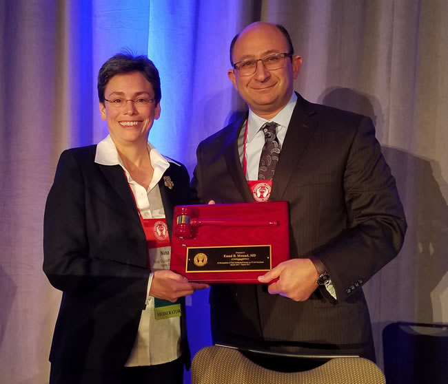
New CCAS president Dr. Nina Guzzetta presents award to Dr. Emad Mossad, outgoing CCAS president, for his years of service
Session I: Basic Science and Clinical Application
Program chair Scott G. Walker, MD introduced the first session of the day.
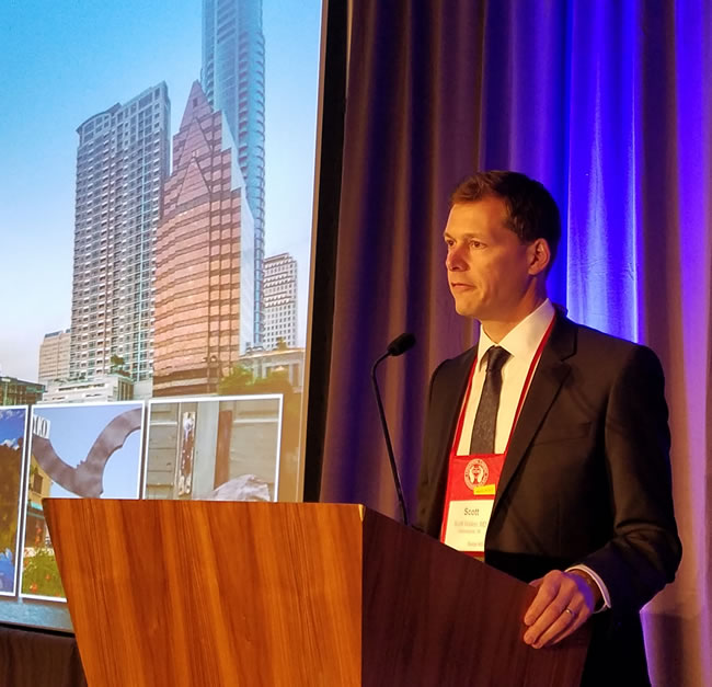
Dr. Scott Walker introduces the CCAS meeting
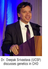
Deepak Srivastava, MD, PhD (Gladstone Institute of Cardiovascular Disease at UCSF, San Francisco) started the session with a talk about applying modern genetic technology to congenital heart disease. The NHLBI consortium collects blood and tissue from thousands of children with CHD as well as their parents, and has developed a database used for sequencing. Multiple genes have been identified related to cardiogenesis, for example GATA4, which functions to regulate atrial and ventricular septation. iPS-derived cardiomyocytes have been developed from fibroblasts or blood cells to behave similarly to pluripotent stem cells. These cardiomyocytes can be altered to have specific point mutations associated with CHD and compared to isogenic control cells.
 Dr. Srivastava’s lab demonstrated that a GATA4 defect led to downregulation of regulator signal Shh in the pulmonary endoderm, which is critical to septation in the neighboring myocardium. Future studies aim to identify drugs and therapies targeted to specific genetic mutations. Another topic of interest for Dr. Srivastava is whether developmental pathways can be used for regeneration of cardiomyocytes. Using specific genes and chemical inhibitors in vitro, researchers have been able to regenerate cardiomyocytes from fibroblasts. This pathway is currently being tested in pigs, with the goal that ischemic scar can be regenerated into functional myocardium.
Cardiac surgeon Mark Rodefeld, MD (Riley Hospital for Children, Indianapolis) followed with a discussion on failing Fontan circulation and his development of a cavopulmonary assist device (CPAD) as a possible future therapy. Single ventricle physiology results in Fontan palliation, which is highly inefficient due to the lack of a subpulmonic ventricle, ultimately culminating in failure. Dr. Rodefeld asserts that the Fontan circulation is a “man-made form of chronic heart failure” that involves all organ systems.
Many of these complications occur despite good ventricular function; in fact, systolic dysfunction does not typically occur until end-stage disease at which point it may be too late to intervene. Transplantation and VAD are currently the only options, each of which carries its own challenges. The CPAD is a subpulmonic device that could be used to restore normal pressures such that a single ventricle patient could be managed with two ventricle physiology. The device is a spinning disc, roughly the size of a dime, in the center of the total cavopulmonary connection that generates a small increase in pressure to maintain preload. It is powered by a biconical motor in the center of the device, developed in conjunction with NASA engineers. Even if the pump stops rotating, it will act as a passive flow diverter which may be better than a completely unsupported cavopulmonary flow. Under these improved conditions, a functioning ventricle could potentially last 30-40 years.
Questions for Session I:
All questions were asked anonymously via the “Ask the Speaker” app feature.
- For Dr. Srivastava: How early can errors causing septal defects be detected and can something be done about it?
In a 7-week fetus most of the morphologic development is done, so it is unlikely to be able to impact the initial morphologic event. The potential is in developing preventive measures or impacting ongoing sequelae. - For Dr. Rodefeld: Will the CPAD require anticoagulation? What about RBC lysis and trauma?
Yes, it will require Coumadin, particularly since it is placed in the venous circulation. The pump will require rigorous thrombogenicity testing in animals before its use.
As for RBC trauma, shear stress can be modulated by engineering. Stasis is the other end of the spectrum that could be a concern, so the pump mechanics have to be just right. Hemolysis rates in cows have been low upon testing thus far. - For Dr. Srivastava: Are there any unintended consequences of switching fibroblasts to myocytes?
Creating pockets of myocardium separated by scar can be a nidus for arrhythmia, which he is concerned about. Currently the process is not efficient enough to cause hypertrophy from overconversion of cells. - For Dr. Rodefeld: What about the efficiency of the pump? What about exercise tolerance?
The pump is quite efficient, but the mechanics are complicated so he leaves that to the engineers. He does not believe the rotation speed will have to be increased with activity. - For Dr. Rodefeld: Will there be in vivo or animal model testing for the CPAD soon?
There is no animal model of a Fontan. Trials in calf or sheep with physiology as a venous loop will have to be the surrogate model. The biggest barrier to commercialization will be small market size. A device for chronic use will have a better chance at commercialization than a short-term device. - For Dr. Srivastava: Do you see your work applying to Dr. Rodefeld’s single ventricle population?
He would like to understand why the RV develops so many problems over time. Perhaps future research could reprogram the RV myocyte to behave more like an LV myocyte. iPS-derived cardiomyocytes are not forceful enough currently to become an LV, but if only 2-3 mmHg are needed, he could imagine a band of muscle around the SVC and IVC which could act as a pump for the Fontan circulation.
Session II: Practice Updates in Congenital Cardiac Anesthesia
Program co-chair Kirsten Odegard, MD moderated an interesting discussion of practice updates in our field.
Dean B. Andropoulos, MD MHCM (Texas Children’s Hospital, Houston) provided the latest information on the second year advanced fellowship programs in congenital cardiac anesthesia. The first recommendations for such training programs debuted in 2010. Currently, there are 58 pediatric anesthesia fellowship programs. For the 2016-17 training year, there are 17 congenital cardiac anesthesia fellowship programs, with two additional programs offering positions next year. Of these programs, nine offer instructorships, and eight have no associated attending duties. Forty percent of the positions went unfilled across the country for 2016-17. According to a PALC survey, the need for congenital cardiac anesthesiologists was nine in 2015, eight in 2016, and five in 2017, thus supply and demand must be considered. It is recommended that programs offer goals and objectives, evaluations, and a robust didactic schedule to the trainees. Boston Children’s Hospital suggests a minimum of 200 cases, with 60% operating room and 40% cath lab. In the future, these programs need to be assessed and promoted by the CCAS and PALC. ACGME accreditation is something to be considered. Program size is not a barrier to this, but there may be advantages to remaining unaccredited, such as the opportunity to utilize the instructorship structure.
Annette Y. Schure, MD (Boston Children’s Hospital, Boston) provided a very informative update on anesthesia for cardiac transplant patients. Median survival of transplant patients is greater than 20 years for infants and greater than 12 years for teens. Improvement in survival is due to advancements in surveillance and immunosuppressive therapies. Re-transplantation occurs in 6% of patients and is associated with worse outcomes. Cardiac transplant patients pose several anesthetic challenges related to physiology, immunosuppression, rejection episodes, and more. With regard to transplant physiology, denervation has multiple consequences, including impaired stress response, reduced exercise capacity, and diastolic dysfunction. The stress response becomes highly preload dependent. There is also an altered response to medications. There is a decreased response to indirect-acting sympathomimetics (dopamine, ephedrine), and little if any heart rate response to atropine or glycopyrrolate. Direct-acting sympathomimetics (epinephrine, norepinephrine), calcium-channel blockers, beta blockers, and adenosine cause an exacerbated response, and dosing may need to be reduced. Reinnervation is possible but patchy, and becomes less likely with each rejection episode. Immunosuppressive medications can have multiple side effects that mimic rejection symptoms. Strict surveillance of transplant patients requires multiple cath procedures. The rate of adverse events is 3.3%, with 1.1% being of high severity. The addition of coronary angiography triples the risk. Risk factors for complications include nonelective procedure, fewer prior caths, longer duration, and low volume centers. The choice of anesthetic for cardiac biopsy is center-specific, but greater than 70% are done with sedation, and greater than 50% are done with spontaneous ventilation. Interestingly, use of mechanical ventilation triples the risk of high severity complications. Whether undergoing cath or noncardiac surgery, the anesthesiologist should ask the following questions:
- What was the indication for transplant?
- Time since transplant?
- Previous caths/biopsies?
- Current functional status?
- Current immunosuppression?
- Elective or emergent?
The use of neuromuscular blockade reversal has come into question in cardiac transplant patients due to case reports of associated cardiac arrest. A more recent series from Mayo Clinic with no episodes of arrests suggests that it may not be the reversal drug itself but rather the patient’s underlying condition. Other medication considerations include suppression of tacrolimus metabolism by high dose dexmedetomidine, which can lead to potential neurotoxicity. A thorough history and physical examination are critical in these patients.
Nina Deutsch, MD (Children’s National Medical Center, Washington DC) followed with an exciting new innovation in the cath lab—MRI-guided right heart cath (RHC). Traditional cardiac catheterization is associated with high levels of radiation exposure. A diagnostic cath has radiation equivalent to 350 chest X-rays, while a complex cath can have levels equivalent to up to 4,000 chest X-rays! MRI-guided RHC is radiation free. This procedure is currently only being done at Children’s National Medical Center, and is being studied in conjunction with the NIH. Non-complex cases older than five years old are eligible. To date, 42 patients have undergone MRI-guided RHC. The most common procedure is diagnostic cath with biopsy, but it has also been used for ASD and PDA device closures. No adverse events or complications have been reported. The major issue thus far is time. On average, there is an extra 34 minutes of scanning to determine which images are best, five minutes of real-time planning, and five to six minutes of research scans. The cath procedure itself takes the same amount of time as usual. Anesthetic implications for MRI-guided RHC include rigorous setup, MRI safety, and resource utilization. Future applications may include EP studies or more complex single ventricle patients.
Questions for Session II:
All questions were again asked anonymously via the “Ask the Speaker” app feature.
- For Dr. Andropoulos: Are there advantages to ACGME accreditation for the congenital cardiac anesthesia fellowship?
He has not looked further into accreditation, just noted it as something to be discussed among the CCAS. - For Dr. Andropoulos: If only 8-10 fellows get a job, but we train many more, will we have a surplus?
This is a topic to be discussed for pediatric anesthesia fellowships in general. We in fact may be training too many fellows. This has not been studied to date, and perhaps we should survey it. - For Dr. Schure: Should we be using sugammadex rather than neostigmine in cardiac transplant patients?
Dr. Schure does not believe neostigmine to be the cause of cardiac arrest in these patients, but rather the underlying disease or rejection episode. - For Dr. Schure: What about dexmedetomidine in cardiac transplant patients?
There is one case report of cardiac arrest with dexmedetomidine and cardiac biopsy. This is more likely related to reduced metabolism of tacrolimus with dexmedetomidine use. Beware of using it in high doses, and adjust dose in neonates and infants. - For Dr. Andropoulos: Is it worth the extra $200,000 it costs a fellow to train an extra year?
Yes, it is. A 12-month fellowship is now required for a position at many academic medical centers. Dr. Odegard agreed, stating that the field is getting more and more advanced.
Session III: Focus on a Lesion: Congenitally Corrected Transposition of the Great Arteries
Program co-chair Luis Zabala, MD moderated the final session of morning, which offered both anesthetic and surgical perspectives on congenitally corrected transposition of the great arteries (cc-TGA).
Premal Trivedi, MD (Texas Children’s Hospital, Houston) provided a thorough overview on the perioperative management of cc-TGA. He began with two audience live polls demonstrating the rarity of cc-TGA. Cc-TGA accounts for 0.5-1% of all CHD. It is characterized by both atrioventricular (AV) and ventriculoarterial (VA) discordance. Situs solitus refers to normal atrial arrangement while situs inversus refers to mirror-image atrial arrangement.
While this anatomy is physiologically corrected, the RV is the systemic ventricle and the tricuspid valve (TV) is the systemic AV valve. Over time, the right heart will fail and the tricuspid valve can become incompetent. There is also potential for abnormal conduction due to anterior and superior displacement of the AV node. The clinical presentation of cc-TGA is variable and depends on associated abnormalities. Those with isolated cc-TGA may be asymptomatic into adulthood, while those with associated pulmonary stenosis or pulmonary atresia may be cyanotic. Intervention depends on the nature of the lesion and severity of symptoms. If there are no associated lesions, and the RV and TV are normal, management is expectant with regular surveillance. Intervention for cc-TGA with associated lesions and/or RV or TV dysfunction varies from single-ventricle palliation, to biventricular repair, to intermediate procedures, to transplantation.
Options for biventricular repair are either physiologic repair or anatomic repair. Historically physiologic repair was performed, but was problematic in that the RV remained the systemic ventricle. Anatomic repair is now favored but requires LV conditioning. This can be done by serial PA banding with gradual tightening over time leading to progressive increase in LV afterload. This is more successful if done at a younger age. Multiple sutures lines, baffle leak or stenosis, and LV dysfunction are concerns associated with anatomic repair. The anesthetic approach to cc-TGA is dependent on each individual patient’s physiology. The acronym CC-TGA can be used as a preoperative checklist for patients with cc-TGA: Conduction, Contractility, Tract obstruction, Gap (VSD), and AV Valve.
Cardiac surgeon Joseph Forbess, MD (Advocate Children’s Hospital, Oak Lawn) led the discussion on surgical management of cc-TGA. He again emphasized right heart failure, LVOT obstruction, and conduction abnormalities as concerns with cc-TGA. How do you correct what is already corrected? The goal is to have the morphologic LV as the systemic ventricle. This implies an atrial inversion and arterial switch or Rastelli. Rastelli-based procedures are options for cc-TGA with associated VSD and PS. In cc-TGA with no associated lesions, the morphologic LV deconditioning dictates the surgical options. Neonatal preparatory PA band followed by double switch operation (DSO) may be best, however early diagnosis may be uncommon. Other options include destination PA band, preparatory PA band with ASD creation, or prophylactic PA band in infancy. LV dysfunction is the major concern in all these surgical options, and cardiac transplantation may loom as the eventual repair.
Questions for Session III:
For Dr. Forbess: How long does the morphologic LV need to be trained?
He is not certain. Patients will not tolerate too tight initial PA band. Some centers use MRI to help determine LV mass, but he has not used this technique. It is variable, as the younger they are, the more rapidly they condition.
- For Dr. Forbess: Have you ever had to do a BT shunt to prevent hypoxia?
No. - For Dr. Trivedi: What about risk stratification for patients with cc-TGA? Should the case be done by cardiac anesthesia?
Regardless of their underlying abnormality, they should be done by cardiac anesthesia more often than not given their unique anesthetic implications. The exception could be made for a patient with cc-TGA/no VSD who is asymptomatic and well followed. - For Dr. Forbess: How do you go about lesion detection?
This is operator dependent. One tip-off is AV valve regurgitation and parallel great arteries. - For Dr. Forbess: With severe TR in these patients, do you perform valvuloplasty or valve replacement?
After DSO, regurgitation greatly improves. If it is noted during the DSO, there is no need to intervene at all. If it is noted later in life, then a valve replacement should be done. - For Dr. Forbess: What do you quote parents in terms of outcome for true cc-TGA?
He does not convince parents to choose the neonatal DSO unless he thinks they will not survive the usual 30-40 years without intervention. He also adds a layer of cardiology counseling between he and the parents.


