


Point of Care Lung Ultrasound: The Stethoscope of the Future?
By Faith J. Ross, MD and Denise Joffe, MD
Introduction
As ultrasound technology has improved over the past few decades, point of care ultrasound has become an increasingly useful tool for anesthesiologists. Most anesthesiologists have adopted ultrasound use for central venous catheter placement and many are facile with transesophageal echocardiography.1
The lungs have traditionally been considered off-limits to ultrasound due to the unfavorable ultrasound acoustics of air-filled organs. Diagnosis of pulmonary pathology such as pleural effusion and pneumothorax historically relied on imaging modalities that utilize ionizing radiation, such as chest radiography and computed tomography. With improvement in technology and training, lung ultrasound has become a useful adjunct to the physical exam and can provide vital information quickly and accurately. In fact, ultrasound can rule out pneumothorax more rapidly and more accurately than chest radiography.2 Ultrasound has greater than 90% sensitivity and specificity for detection of pneumothorax, pleural effusion, and alveolar interstitial disease.2,3
Thoracic ultrasound is increasingly utilized in critical care, emergency medicine, trauma surgery, internal medicine, pediatrics, and anesthesiology.1,4 This review will discuss techniques for basic lung ultrasound and describe the ultrasound appearance of common pulmonary pathology such as pneumothorax, pleural effusion, pulmonary edema, and pneumonia.
The Normal Lung Exam
Ultrasound waves produced by the transducer probe are transmitted through or reflected from tissue planes depending on the acoustic properties of the tissue. Reflected waves are received by the transducer and transformed into an electrical signal to form an image. Ultrasound waves penetrate solid organs and fluid well, but are reflected by air and bone.1,5 As ultrasound tissue penetration depends on the frequency of the transmitted waves, appropriate probe choice should consider the depth of the structure of interest. Imaging of the pleura can be accomplished with a higher frequency (7.5-10 MHz) probe, but deeper structures require a lower frequency (3.5 MHz) probe.5 Both standard 2D imaging and M-mode, which displays the motion of one part of the image over time, are useful for evaluation of pulmonary pathology.
A thorough thoracic ultrasound examination evaluates each thoracic zone (Figure 1) with the transducer perpendicular to the axis of the ribs to obtain an image similar to the one shown in Figure 2. The bright linear structure seen between the rib shadows represents the pleural interface. Movement of the lung with respiration causes a sliding motion of the visceral pleura over the parietal pleura referred to as lung sliding. In normal lung, reflection of ultrasound waves from the pleural interface creates a series of equally spaced horizontal lines referred to as A lines. Repetitive reflection of ultrasound waves within in the lung parenchyma creates vertical B lines (also sometimes called comet-tail artifact or lung rockets), perpendicular to the pleural line, which extend from the pleura to the bottom of the image and obliterate transecting A lines.6 A normal lung exam should reveal lung sliding, A lines, and fewer than three B lines between a pair of ribs.6
In M-mode, the normal lung appearance is described as a “seashore,” with the overlying soft tissues representing the waves, and the underlying mobile pleura and parenchyma as the sandy beach. (Figure 3) The lung pulse refers to a small movement of the pleura with cardiac pulsation. It should not be confused with lung sliding which bares no relationship to the cardiac cycle.7 Lung pulse is subtle and typically not appreciated unless respiratory motion of the lung is abolished. Pathologic changes in the fluid content or aeration of the lungs produces changes in this typical ultrasound appearance.
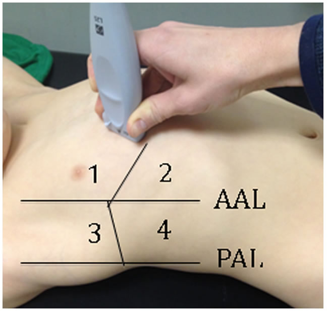
Figure 1: Thoracic zones for complete lung ultrasound examination
AAL: Anterior axillary line, PAL: Posterior axillary line
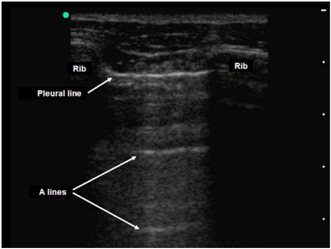
Figure 2: Normal thoracic ultrasound demonstrating pleural line, A lines, and rib shadows.13
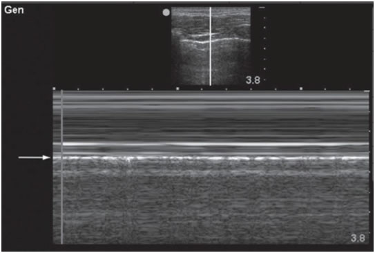
Figure 3: M-mode appearance of normal lung with seashore sign.14
Pneumothorax
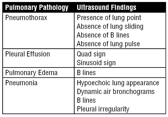
Pneumothorax is characterized by absence of lung sliding, as the interface between the visceral and parietal pleura is no longer present. Lung sliding may also be absent in other pathologies that involve nonaeration of the lung, including endobronchial intubation, severe ARDS, or atelectasis. As the ultrasound transducer is moved to an area of aerated lung, lung sliding should become visible. This “lung point” is a specific diagnostic finding for pneumothorax.8 In M-mode, the typical seashore appearance is replaced by a static “barcode” appearance. The transition between the “seashore” and “barcode” areas of the lung identifies the lung point. (Figure 4) The presence of A lines does not rule out pneumothorax as they are sometimes present even in the absence of aerated lung.5 B lines are not present in pneumothorax. If a lung point cannot be identified, but lung sliding, B lines, and lung pulse are absent, alternative methods should be used to verify the diagnosis of pneumothorax.

Figure 4: Appearance of lung point in 2D and M-mode ultrasound. The arrow denotes the lung-point. Note the transition from “seashore” to “barcode” in the right portion of the image.6
Pleural Effusion
Pleural effusion should be evaluated with the ultrasound probe placed as posteriorly as possible in a supine patient to identify fluid layered in the dependent portion of the thorax. Effusion appears as an anechoic (black) area deep to the parietal pleura referred to as the quad sign. The upper boundary of the area is created by the parietal pleura and the lower boundary by the visceral pleura. Movement of the lung toward the pleural line on inspiration creates an alternating pattern on M-mode known as sinusoid sign.6,9 (Figure 5)
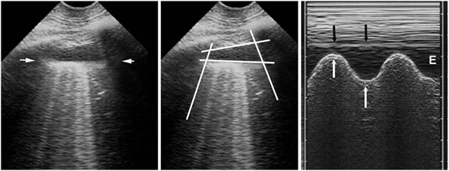
Figure 5: Appearance of pleural effusion in 2D and M-mode ultrasound. Note the quad sign in the middle image and the M-mode sinusoid sign in rightmost image.6
Pulmonary Edema
Pulmonary edema is characterized by multiple B lines created by repetitive echo reflection from thickened and edematous interlobular septa.6 (Figure 6) B lines may be seen in acute pulmonary edema, acute respiratory distress syndrome, pneumonia, or chronic interstitial lung disease.1 B lines change acutely with patient condition, increasing in number with fluid loading and decreasing with fluid removal during hemodialysis.10 It may be possible to use dynamic changes in B lines to assess volume resuscitation and adequacy of diuresis.
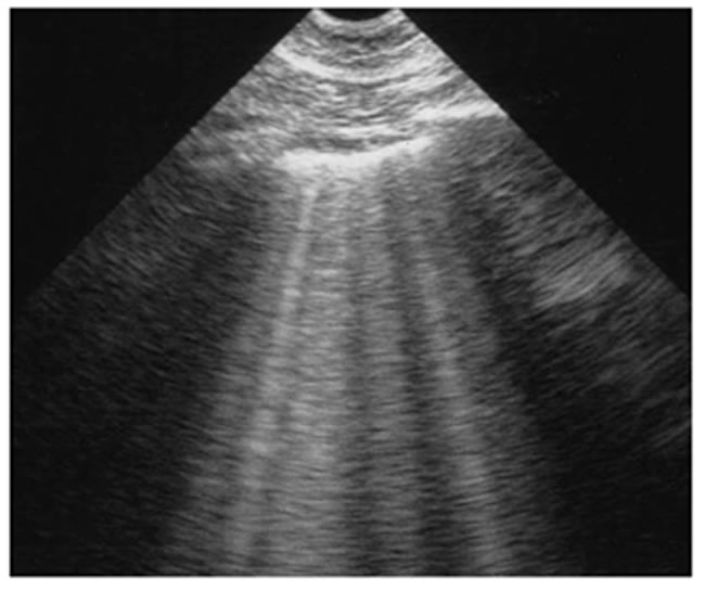
Figure 6: Multiple B lines suggestive of interstitial/alveolar fluid. Note that the B lines begin at the pleura and extend to the bottom of the image. A lines cannot be seen crossing the B lines.6
Pneumonia
Consolidated lung takes on a hypoechoic appearance similar to fluid rich tissues such as the liver or spleen. Pneumonia is differentiated from other causes of consolidation by dynamic air bronchograms, visualized as bright linear structures converging toward the hilum.11 (Figure 7) Air entrapment inside atelectatic lung may cause static air bronchograms that do not change with respiratory motion. Lung ultrasound is superior to chest x-ray in detecting pneumonia; however, sonographic diagnosis of pneumonia is limited to subpleural regions of the lung that can be accessed without sonographic interference from overlying aerated lung. Other ultrasound characteristics of pneumonia include B-lines, pleural irregularity, and lung sliding with or without A-lines.
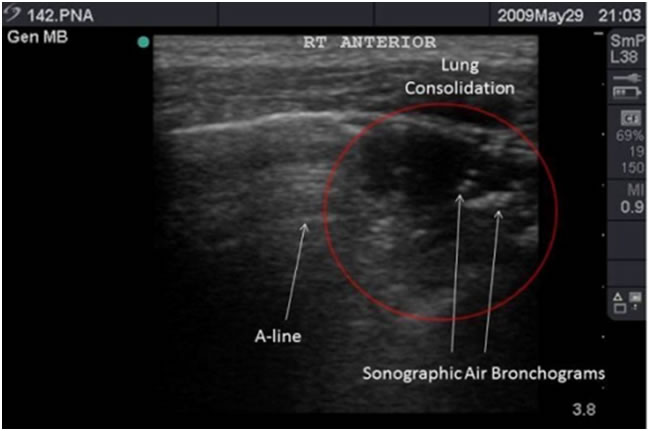
Figure 7: Sonographic air bronchograms in a patient with an ultrasound diagnosis of pneumonia.15
Conclusion
Point of care lung ultrasound has become a useful tool for quickly evaluating pulmonary pathology in real time and can be a valuable resource for anesthesiologists in the operating room or intensive care unit. As portable ultrasound technology progresses in the future, ultrasound evaluation of the heart and lungs may become a routine extension of the physical exam. We strongly recommend that all anesthesiologists familiarize themselves with this incredibly useful technique.
References
- Moore CL, Copel JA. Point-of-Care Ultrasonography. N Engl J Med. 2011;364(8):749–757. doi:10.1056/NEJMra0909487.
- Blaivas M, Lyon M, Duggal S. A prospective comparison of supine chest radiography and bedside ultrasound for the diagnosis of traumatic pneumothorax. Acad Emerg Med. 2005;12(9):844–849. doi:10.1197/j.aem.2005.05.005.
- Lichtenstein D, Goldstein I, Mourgeon E, Cluzel P, Grenier P, Rouby J-J. Comparative diagnostic performances of auscultation, chest radiography, and lung ultrasonography in acute respiratory distress syndrome. Anesthesiology. 2004;100(1):9–15.
- Kirkpatrick AW, Melniker L, Gargani L, Noble VE. International evidence-based recommendations for point-of-care lung ultrasound - Springer. Intensive care …. 2012.
- STEFANIDIS K, DIMOPOULOS S, NANAS S. Basic principles and current applications of lung ultrasonography in the intensive care unit. Respirology. 2011;16(2):249–256. doi:10.1111/j.1440-1843.2010.01885.x.
- Lichtenstein DA. Lung ultrasound in the critically ill. Ann Intensive Care. 2014.
- Piette E, Daoust R, Denault A. Basic concepts in the use of thoracic and lung ultrasound. Current Opinion in Anaesthesiology. 2013;26(1):20–30. doi:10.1097/ACO.0b013e32835afd40.
- Lichtenstein D, Mezière G, Biderman P, Gepner A. The “lung point”: an ultrasound sign specific to pneumothorax. Intensive Care Med. 2014;26(10):1434–1440. doi:10.1007/s001340000627.
- Lichtenstein DA. Ultrasound examination of the lungs in the intensive care unit. Pediatric Critical Care Medicine. 2009;10(6):693–698. doi:10.1097/PCC.0b013e3181b7f637.
- Noble VE, Murray AF, Capp R, Sylvia-Reardon MH, Steele DJR, Liteplo A. Ultrasound Assessment for Extravascular Lung Water in Patients Undergoing Hemodialysis. Chest. 2009;135(6):1433–1439. doi:10.1378/chest.08-1811.
- Weinberg B, Diakoumakis EE, Kass EG, Seife B, Zvi ZB. The air bronchogram: sonographic demonstration. AJR Am J Roentgenol. 1986;147(3):593–595. doi:10.2214/ajr.147.3.593.
- Gargani L, Volpicelli G. How I do it: Lung ultrasound. Cardiovascular Ultrasound 2014 12:1. 2014;12(1):1. doi:10.1186/1476-7120-12-25.
- Ioos V, Galbois A, Chalumeau-Lemoine L, Guidet B, Maury E, Hejblum G. An integrated approach for prescribing fewer chest x-rays in the ICU. Ann Intensive Care. 2011;1(1):1. doi:10.1186/2110-5820-1-4.
- Stone MB. Ultrasound diagnosis of traumatic pneumothorax. J Emerg Trauma Shock. 2008;1(1):19–20. doi:10.4103/0974-2700.41788.
- Tsung JW, Kessler DO, Shah VP. Prospective application of clinician-performed lung ultrasonography during the 2009 H1N1 influenza A pandemic: distinguishing viral from bacterial pneumonia. Crit Ultrasound J. 2012;4(1):16. doi:10.1186/2036-7902-4-16.
Re-printed with permission from the American Academy of Pediatrics Section on Anesthesiology and Pain Medicine newsletter.


