


 Can’t ventilate? Is it severe bronchospasm or something else?
By Peggy P McNaull, MD and Sara L Pittenger, MD
North Carolina’s Childrens Hospital, Chapel Hill, North Carolina
Case:
The patient is a former 35 week gestation infant delivered prematurely secondary to uterine rupture. Following birth, he was observed overnight in the neonatal intensive care unit and then discharged home from the hospital on day of life 5 in stable condition. On day of life 12, he presented to an outside institution with bilious emesis and, subsequently, underwent an exploratory laparotomy and an open repair of duodenal stenosis. The operative course was relatively uneventful with the exception that intubation took several attempts and refluxed milk was noted in the posterior pharynx. There was no intraoperative hypoxia. Nonetheless, the patient remained intubated and ventilated postoperatively in the pediatric intensive care unit (PICU) secondary to concerns for the patient’s young age, possible intraoperative aspiration, and significant postoperative pain.
In the evening of postoperative day (POD) 0, the patient was hypotensive and required both significant inotropic and ventilatory support. The PICU team presumed the patient’s symptomatology was secondary to systemic inflammatory response syndrome (SIRS) and aspiration pneumonitis. On POD 1, the patient became acutely hypoxic and hypercarbic and ultimately suffered a cardiac arrest.
Several attempts at reintubation were made during the resuscitation and despite visualization of the endotracheal tube through the glottic opening breath sounds were not appreciated. After 40 minutes of resuscitation, there was return of spontaneous circulation and breath sounds were appreciated at high peak inspiratory pressures through a 3.0 cuffed endotracheal tube (ETT). The PICU team noted that the exact cause of the cardiopulmonary arrest was unclear but likely attributable to bronchospasm, plugging, or acute respiratory distress syndrome (ARDS). A post resuscitation echocardiogram demonstrated normal cardiac function with a patent ductus arteriosus and a patent foramen ovale. An MRI of the brain obtained several days after the cardiopulmonary arrest demonstrated changes consistent with a significant hypoxic/anoxic injury.
Over the next 10 days, the patient was intermittently difficult to ventilate. A tracheal aspirate demonstrated methicillin resistant staphylococcus aureus. Antibiotics, albuterol, solumedrol, and heliox were all used with varying amounts of success. On POD 10, otolaryngology was consulted and flexible bronchoscopy demonstrated an area of soft tissue at the distal tip of the endotracheal tube. It was unclear whether this was granulation tissue, some form of stenosis, or due to compression from an external source. The patient was then transported to our institution for further evaluation by our pediatric airway center.
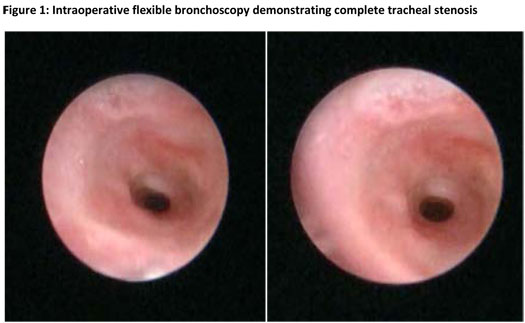
Flexible bronchoscopy with 2.2 mm bronchoscope revealed at least three to four complete tracheal rings with the distal ring located just above the carina (Fig. 1). The stenotic area was estimated to be approximately 2.5 mm in diameter. Other pertinent findings observed with bronchoscopy included a tracheal bronchus and abnormal left-sided bronchial anatomy with no left mainstem bronchus.
The patient presented to the operating room intubated with a 3.0 cuffed ETT secured at 8 cm for a slide tracheoplasty on cardiopulmonary bypass (CPB). Prior to the initiation of CPB, the patient had several episodes of hypoxia and inadequate ventilation that resolved with gentle ETT repositioning and hand ventilation. Surgical examination aided by flexible bronchoscopy demonstrated that the area of stenosis occupied at least 50% of the trachea. Following surgical repair and prior to separation from CPB, positive pressure ventilation was applied up to 35 cm of water pressure to ensure no tracheal leak and the ETT was positioned below the level of the proximal anastomosis. The patient separated from CPB without incident and was transferred to the PICU in stable condition.
The infant had a protracted postoperative course. He returned to the operating room on one occasion for laser excision of granulation tissue. Ultimately, his neurologic status necessitated the placement of both a tracheostomy and gastrostomy tube. He was discharged home with his family approximately 3 months after his initial presentation with duodenal stenosis.
Discussion:
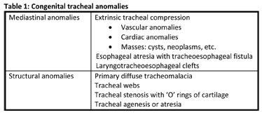
Congenital tracheal anomalies occur in an estimated 1 in 60,000 live births and are a result of either extrinsic mediastinal anomalies or structural anomalies of the trachea itself (Table 1).
Congenital tracheal stenosis (CTS), a rare but life-threatening disorder, is an anomaly of the trachea’s cartilaginous skeleton characterized by the presence of complete, circular ‘O’ rings of cartilage (absent posterior pars membrosa). The complete tracheal rings vary in location, severity, and length. A classification system of four different types has been described by Grillo (Fig. 2).

Type I: long-segment stenosis involving almost the entire trachea Type II: funnel shaped trachea of various locations and variable lengths Type III: short-segment stenosis with or without anomalous right upper lobe bronchus Type IV: bridge bronchus originating from a pseudo-carina created by the tracheal origin of the right upper lobe bronchus and connecting the trachea via horizontally branching bronchi to the rest of the lungs
There is also a high incidence of other cardiovascular, respiratory, and/or gastrointestinal anomalies; most frequently a left pulmonary artery sling and/or a tracheal origin of the right upper lobe bronchus. Patients typically present within the first few weeks to months of life with biphasic stridor. The length of involved trachea is less critical than the degree of stenosis in producing symptoms. Airway resistance increases fourfold as the luminal diameter decreases. In fact, symptoms may not become apparent until the onset of an acute upper respiratory infection that results in airway edema and a subsequent reduction in tracheal diameter.
As a result of its rare occurrence, experience in the diagnosis and management of patients with congenital tracheal stenosis is limited. Consequently, the diagnosis of CTS is often delayed. As detailed above, a delay in diagnosis can lead to devastating results. Unanticipated difficult intubation in an infant without obvious craniofacial abnormalities is a rare event. When the intubation of a neonate or infant is difficult, CTS must be suspected and the patient referred to a center experienced in the care of this complex disease.
In a patient with CTS, the slightest mucosal injury caused by repeated intubation attempts, bronchoscopy, or even a URI may lead to clinical decompensation and an acute airway crisis for which emergent tracheosotomy is potentially of no benefit. In the event of a difficult intubation or an inability to ventilate despite apparent tracheal intubation, the placement of an LMA should be considered and has been used with success in children with CTS.
The management of the patient with CTS mandates a multidisciplinary approach with the participation of pediatric general surgeons, cardiovascular surgeons, otolaryngologists, anesthesiologists, and pulmonologists. It is proposed that the pediatric patient can tolerate up to 50% narrowing of the trachea before respiratory symptoms become apparent. Hence, many suggest that surgery is only indicated in the setting of severe respiratory symptoms and/or a stenotic segment less than 40% of the normal tracheal diameter.
Many surgical approaches have been used in the treatment of CTS including resection with end-to-end anastomosis, enlargement patch tracheoplasties, tracheal autografts or homografts, and slide tracheoplasty. Among these techniques, slide tracheoplasty has emerged as the best surgical option for the treatment of long segment stenosis (Fig. 3).

(a) The stenotic segment is transected exactly at its midpoint. (b) The stenotic segment is longitudinally slit anteriorly on its caudal segment and posteriorly on its cranial segment. (c) By gentle traction, the upper and lower segments are slid over one another. (d) An oval-shaped, oblique from posterior to anterior anastomosis is made, thereby shortening the trachea by half and increasing its cross-sectional area by four
Prior to the 1980s, severe CTS was associated with high mortality rates. As surgical advancements have been made, outcomes for patients with CTS have dramatically improved. Regardless of improvements in surgical technique, the early recognition and diagnosis of CTS is critical to long term outcome and survival. Albeit rare, the anesthesiologist must be familiar with CTS and have a high index of suspicion in the event of a difficult intubation in a neonate or infant.
References:
Ali MI, Brunson CD, Mayhew JF. Failed intubation secondary to complete tracheal rings: a case report and literature review. Pediatric Anesthesia 2005; 15: 890-92.
Ho AS, Koltai PJ. Pediatric Tracheal Stenosis. Otolaryngol Clin N Am 2008; 41: 999-1021.
Monnier P (ed). Pediatric Airway Surgery: Management of laryngotracheal stenosis in infants and children. 1st edition. New York: Springer; 2011.
Terada M, Hotoda K, Toma M, et al. Surgical management of congenital tracheal stenosis. Gen Thorac Cardiovasc Surg 2009; 57: 175-83.
Figures included with kind permission from Springer Science+Business Media: Pediatric Airway Surgery: Management of laryngotracheal stenosis in infants and children, 2011, pg. 169 & 173, P Monnier, figure numbers 13.11 and 13.15.
