


Image of Interest
Authors
Christopher D. Denny, MD and Pablo Motta, MD, FAAP
Baylor College of Medicine and Texas Children’s Hospital
Keywords
Central veins, MR venography, venous imaging
Case Presentation
A 17-year-old male presents with three days of right arm pain and substernal chest pain. His past medical history is significant for diffuse midline glioma status post-resection. He has a left subclavian port-a-cath for chemotherapy and laboratory blood draws. The physical exam is significant for edema in the right arm painful to motion and blanching erythema across the upper chest.
Chest x-ray shows port-a-cath with the tip in the superior vena cava (SVC) and right atrium (RA) junction (Figure 1A). Transesophageal echocardiographic (TEE) examination is suspicious for port-a-cath thrombosis with almost total occlusion of the superior vena cava and thrombus extension into the right atrium (Figure 1B).
To confirm the diagnosis, magnetic resonance venography (MRV) was ordered showing thrombosis of the entire right internal jugular vein beginning at the level of the supraglottic larynx to the thoracic inlet with the absence of contrast enhancement in the SVC (Figure 1C and 1D).
In the interventional radiology suite, the patient underwent TEE-guided mechanical thrombectomy with recanalization, venoplasty, and removal of port-a-cath. Post-procedure he was placed on intravenous heparin and transitioned to subcutaneous enoxaparin.
On discharge, he was neurologically intact. Anticoagulation was recommended for 12 months.
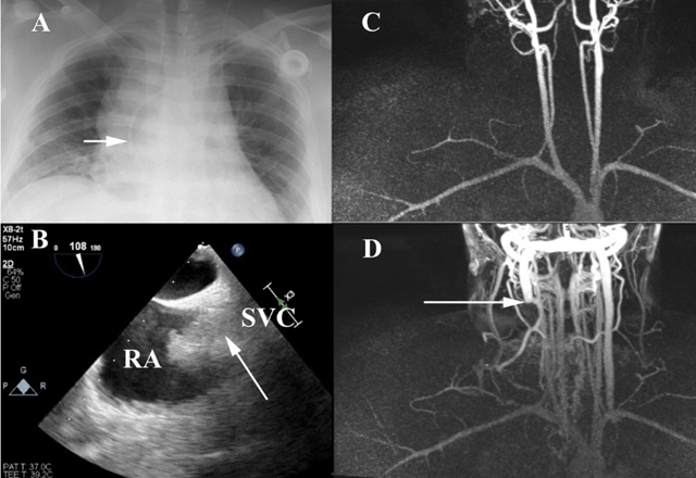
Figure 1: A) Anteroposterior chest x-ray showing the port-a-cath tip at the SVC-RA junction (white arrow); B) Mid esophageal bi-caval view displaying an almost total occlusion of the SVC by thrombus (white arrow); C) MRA normal arterial signal; D) MRV thrombosis of the entire right internal jugular vein beginning at the level of the supraglottic larynx to the thoracic inlet with absence of contrast enhancement in the SVC (white arrow).
Abbreviations: SVC, superior vena cava; RA, right atrium; MRA, magnetic resonance angiography; MRV, magnetic resonance venography.
Reference:
- Cline B, Hurwitz LM, Kim CY. MR Venography of the Central Veins of the Thorax. Top Magn Reson Imaging. 2017; 26(4): 167-174. doi: 10.1097/RMR.0000000000000139. PMID: 28777165.
- Miller A, Schneider J: Pacemaker-Associated Superior Vena Cava Syndrome. N Engl J Med. 2021; 384: e63. DOI: 10.1056/NEJMicm2031975


