


Pediatric Cardiac Magnetic Resonance Imaging
Sujatha Buddhe, MB BS, MS
Division of Pediatric Cardiology
Seattle Children’s Hospital, University of Washington School of Medicine
Brian Soriano, MD
Division of Pediatric Cardiology
Seattle Children’s Hospital, University of Washington School of Medicine
Gregory Latham, MD
Anesthesiology and Pain Medicine
Seattle Children’s Hospital, University of Washington School of Medicine
Cardiac MRI (CMR) is increasingly being used in the diagnosis and management of complex congenital and even acquired heart disease (1, 2). Echocardiography remains the primary imaging modality for the assessment of pediatric heart disease secondary to its easy availability and diagnostic accuracy (3). However, echocardiography has limitations for evaluating extracardiac vascular structures and poor acoustic windows pose problems in some patients particularly for assessment of the right ventricle. CMR is a powerful noninvasive imaging modality which is supplanting cardiac catheterization as the alternative in select situations by providing high quality anatomic and physiologic data including ventricular function and flows (4).
Indications
CMR is indicated in any patient when echocardiography cannot provide all the necessary anatomic or functional information. However, the decision should be based on the risk-benefit profile, particularly in smaller children who would need anesthesia. The indication varies based on institutional practice. Generally in smaller children, where echocardiography provides good intracardiac images with great details, CMR would be indicated to delineate vascular structures such as coarctation and vascular rings, ventricular volumes in borderline hypoplastic ventricles, relationship of complex ventricular septal defects to inflows and outflows, and flows through different vessels to measure shunts. In older children with poor acoustic windows the indications widen further and include assessment of the right ventricle, vascular structures, coronary arteries, cardiomyopathies, tumors and also iron loading in thalassemias (5).
Sequences
Most often, if there are no time or sedation constraints, a full baseline study that includes most of the following sequences is performed. In follow up studies and other situations, fewer sequences are typically used. The commonly used sequences are discussed below:
Cine imaging: The cine images provide multiphasic data averaged over multiple heart beats to assess dynamic function of any structure. For example, long axis or short axis stack from the base to apex of the heart is used to accurately assess ventricular volumes and function (figure 1). They also help assess the dynamic nature of outflow tract obstruction and valvular regurgitation.
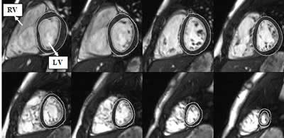
Figure 1:
Short axis steady state free procession (SSFP) image from base to apex of the ventricles. Contours are drawn around the ventricular endocardium and epicardium in end diastole and systole to measure ventricular mass, size and function. LV – left ventricle; RV – right ventricle.
Black blood imaging: These sequences provide two-dimensional images that are particularly useful for the assessment of blood vessel stenosis where turbulent flow reduces accuracy of cine imaging. It is also useful for assessing airway compression in vascular rings and slings.
Flow assessment: Using a technique called phase contrast sequences, flow across blood vessels and valves can be measured to assess regurgitation and shunts (6, 7). The pulmonary to aorta blood flow ratio can help measure Qp:Qs and assess the shunt across an atrial or ventricular septal defect (VSD).
Angiography: 3D angiography can be performed with or without intravenous contrast. However, the signal and quality of the images improve significantly with contrast-based imaging. The time resolved component of this sequence can be used to assess flow path and patterns for the diagnosis of anomalous arterial or venous connections and collateral vessels. The relatively newer blood pool contrast agent “Gadofosveset Trisodium” (Ablavar) allows ECG- and respiratory-gated, free breathing, high spacial resolution angiography images (8, 9).
Late gadolinium enhancement imaging: The delayed or late gadolinium enhancement (LGE) images are other important sequences that are unique to CMR. The diseased myocardium in cardiomyopathies and postoperative patients is enhanced in these sequences (10, 11). Multiple studies have shown that this information helps with risk stratification (12, 13). Along with other sequences, LGE also helps in characterizing cardiac tumors (14).
Stress perfusion imaging: This sequence helps analyze any perfusion defects associated with coronary artery disease such as anomalous coronary origins, Kawasaki disease and surgeries involving coronary artery translocation such as arterial switch in transposition (15). Typically, ventricular perfusion and function are assessed with either adenosine or dobutamine infusion.
Clinical applications
- Coarctation and aortic arch anomalies: CMR is very useful in the assessment of aortic coarctation and arch anomalies such as vascular rings and slings (16-18). Angiography delineates the arch anatomy very well (figure 2) along with collateral vessel assessment. Post stent or surgical intervention, CMR is even more useful for assessment of residual stenosis or post-procedural complications such as aneurysm or dissection. If needed, functional imaging with ventricular mass assessment could be performed to assess hemodynamic significance.
- Tetralogy of Fallot: Tetralogy post repair is one of the most common indications for performing CMR’s in any institution. CMR is the gold standard imaging modality for analyzing right ventricular size and function (19-22). As well as right ventricle evaluation, MR angiography is performed to evaluate branch pulmonary artery and conduit stenosis (Video 1). Flow analysis also provides unique data to analyze pulmonary regurgitant fraction and differential flow through right versus left branch pulmonary arteries (23). Presence of delayed enhancement adds important prognostic information (12, 24)
- Cardiomyopathies: CMR provides unique data in the diagnosis, management and follow up of different cardiomyopathies like hypertrophic, dilated cardiomyopathies and arrhythmogenic right ventricular dysplasia (25). CMR provides accurate quantification of ventricular size, mass, myocardial architecture, and segmental and global function. Delayed enhancement imaging again provides prognostic information (13, 26).
- Complex congenital heart disease: In complex congenital heart diseases such as double outlet right ventricle (DORV) with borderline hypoplastic ventricles, CMR provides valuable information on ventricular volumes to guide the surgical strategy of single versus biventricular repair. Also, ECG-gated isotropic angiography is ideal to assess the three-dimensional relationship of intracardiac structures, such as the VSD and its relationship to inflow and outflow tracts. The latest advancements in 3D printing of heart models utilizing this data provides critical information to the surgeons in preparing for a complex repair of baffling the left ventricle to systemic outflow tract through VSD (figure 3).
- Single ventricle: In patients undergoing palliation via the Fontan pathway, CMR provides unique data in different stages of surgical repair. Anatomic data at each stage helps assess any residual coarctation or branch pulmonary artery stenosis (Figure 4). Some institutions are performing CMR instead of or in addition to cardiac catheterization prior to their Glenn and Fontan surgical repairs (4, 27-29). These studies are performed in greater detail evaluating anatomy, function, residual shunts, stenosis and collateral flow at each stage (30).
- Other indications: Other common lesions where CMR provides unique important information are myocarditis (31), partial anomalous pulmonary venous return (figure 5), anomalous origins of coronary arteries (figure 6) and post repair of any congenital heart disease, such as transposition of great arteries (32).
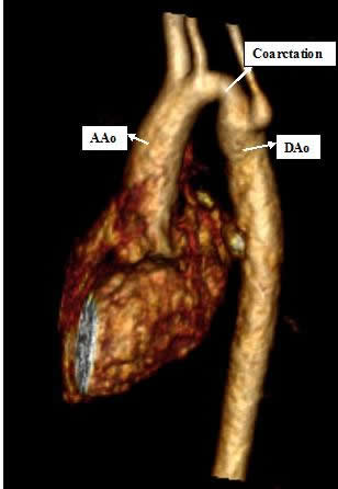
Figure 2:
3D contrast enhanced MR angiogram showing coarctation of aorta with narrowing in the transverse arch. AAo – Ascending aorta; Dao – Descending aorta; Coarctation
Video 1
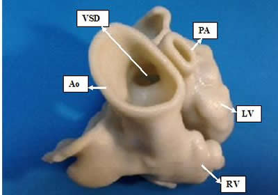
Figure 3:
3D printed heart model based on ECG gated magnetic resonance angiography (MRA) in a patient with transposition of great arteries, ventricular septal defect and pulmonary stenosis. The model is created mainly to help complex repair of baffling the left ventricle to systemic outflow tract through VSD. LV – left ventricle; RV – right ventricle; Ao – Aorta; PA – pulmonary artery; VSD – ventricular septal defect.
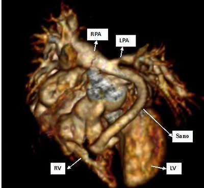
Figure 4:
3D contrast enhanced MR angiogram viewed from left anterior in a 3 month old status post Norwood Sano shunt procedure being evaluated for a biventricular repair. The reconstructed image shows proximal left pulmonary artery stenosis at the insertion of the Sano shunt. RV – right ventricle; LV – left ventricle; Sano – Sano shunt; LPA – left pulmonary artery; RPA – right pulmonary artery.
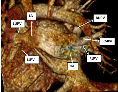
Figure 5:
3D contrast enhanced MR angiogram viewed from posterior in a patient with partial anomalous pulmonary venous return. Three right pulmonary veins are seen draining directly into the right atrium while the left pulmonary veins drain normally into the left atrium. RA – right atrium; LA – left atrium; RUPV – right upper pulmonary vein; RMPV – right middle pulmonary vein; RLPV – right lower pulmonary vein; LUPV – left upper pulmonary vein; LLPV – left lower pulmonary vein.
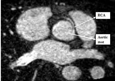
Figure 6:
3D contrast enhanced MR angiogram axial view showing anomalous origin of right coronary artery from left coronary cusp at 3 o’clock position. RCA – right coronary artery; Aortic root.
Contraindications
The primary contraindication for CMR is patients with pacemakers and defibrillators. However, some institutions are beginning to perform occasional CMR’s in patients with pacemakers following strict safety protocols (33, 34). Other relative contraindications include patients with certain types of metal implants (e.g., coils in blood vessels), mainly because of the significant artifact created that degrades image quality. Gadolinium contrast is contraindicated in patients with significant renal dysfunction. Lastly, if sedation or general anesthesia is required to accomplish the scan and anesthesia is deemed high risk, then the benefits of CMR versus CT scan/catheterization must be carefully discussed by a multidisciplinary group prior to the scan.
Considerations in pediatric patients
In children, the body size is small, heart rates are rapid and there is potential for sudden decompensation. The cardiologist/radiologist should be trained to expedite the sequences and the study while still obtaining necessary information in these cases. In addition, an anesthetic team trained to care for children with hemodynamic issues would be needed. A team of cardiologist, radiologist and anesthesiologist working together is of considerable importance in these situations. While breath-holding sequences significantly improve image quality, in uncooperative awake patients or sedated children with natural airways, free breathing averaging techniques can be applied.
Anesthetic considerations
Most infants and young children require sedation or anesthesia for MRI scans. Very few can tolerate the cold, noisy, and claustrophobic environment, coupled with the need for immobility, during relatively long scan times. The MRI scanner is one of the most challenging physical environments in which children are anesthetized. The major challenges include a location that is usually removed from the operating suites and supportive help, movement of the anesthetized child from the induction to scanner area, requirement of unique non-ferrous monitoring and anesthetizing equipment, and difficult visualization and assessment of the patient once in the scanner. It is very important to have trained anesthesia personnel capable of caring for children with complex cardiac lesions who have the potential to develop cardiac compromise during the scan, and many institutions utilize dedicated pediatric cardiac anesthesiologists during CMR.
Rate of adverse events under anesthesia: There are no large studies assessing risk of adverse events in children undergoing anesthesia for standard MRI, and the small studies that exist suffer from lack of comparison due to varying drug regimens and whether anesthesia was provided by anesthesiologists or non-anesthesiologists, making outcomes difficult to combine. In lieu of such large studies, the large data set from Cravero et al (35) can be used as a surrogate of the risk of propofol sedation outside the operating room. Of the nearly 50,000 children who received propofol for sedation/anesthesia outside the operating room, the rate of serious events was rare (no deaths, cardiopulmonary resuscitation twice), and the rate of desaturation greater than 30 seconds was rather uncommon, occurring at a rate of 154 times/10,000 anesthetics. On the other hand, anesthetizing children with heart disease for non-cardiac procedures carries a heightened risk of morbidity and mortality compared to the general population (36, 37), and anesthetizing such children in MRI intuitively combines both the risk of the heart condition and the MRI environment. Studies reporting anesthesia risk for CMR are few and include small numbers of patients.
Odegard et al reported a very low rate of complications during CMR (38). Out of 250 children undergoing general endotracheal anesthesia, no scans were interrupted because of desaturation or hypotension, no patients were admitted postoperatively for anesthesia-related factors, 5 episodes of hypotension were successfully treated with intravenous medications, and 2 ICU patients received dopamine infusion with the induction of anesthesia. Brown et al (4) compared 41 children undergoing cardiac catheterization and 41 children receiving CMR for pre-Glenn evaluation. The rate of minor adverse events (e.g., prolonged desaturation, acidosis, unplanned ICU admission, arrhythmias, initiating inotropes) was significantly higher in the cardiac catheterization group (83%) versus the CMR group (5%). One child in the CMR group required cardiopulmonary resuscitation (secondary to aortopulmonary shunt thrombosis) compared to none in the catheterization group, but this was not statistically significant.
The study by Rangamani et al (39) analyzed the records of 143 infants less than 120 days old who underwent CMR. Most received general anesthesia; the others received deep sedation or swaddling for immobility. One major adverse event occurred, which was respiratory arrest in a sedated neonate and was easily resuscitated. Minor adverse events occurred in 8%, and the adverse events were unrelated to patient age, complexity of heart disease, prostaglandin E1 dependence, or type of anesthesia. They concluded that CMR could be performed safely in neonates and young infants. Stockton et al (40) also demonstrated the relative safety of CMR. The rate of minor adverse events was 28%, yet all were mild and easily treatable. One patient suffered cardiac arrest during the anesthetic but was successfully resuscitated.
Overall, the reported risk of major adverse events during anesthesia for children undergoing CMR is low. The rate of minor and treatable adverse events is also relatively low for this patient cohort and less than that during cardiac catheterization in a similar patient cohort (4).
Anesthesia monitoring: The same level of safety and monitoring must be provided during anesthesia for MRI as is required in the operating room, including standard monitors. However, radiofrequency during MRI can cause interference with monitors, and monitors may likewise degrade MRI image quality (41). Therefore, MRI-compatible monitors and resuscitation equipment must be used. Reliable ECG monitoring is necessary not only for surveillance of cardiac dysrhythmias but also for triggering ECG-gated sequences during CMR. Additionally, patient temperature must be closely monitored as the low temperature in MR scanning room provides a risk of hypothermia, particularly in small infants. In those children with pre-existing invasive arterial blood pressure monitoring, a non-invasive cuff should be placed because radiofrequency interference of the transducer and subsequent unreliable arterial waveform is possible.
Anesthesia technique: Different institutions use varying types of anesthesia and sedation during CMR (4, 38-40, 42, 43). General anesthesia is usually needed in children less than 7 years of age (38, 44), but this should be determined on a case-by-case basis. General endotracheal intubation affords optimal image acquisition and hastens the overall scanning time. General endotracheal anesthesia also allows adequate ventilation to avoid hypoxia and hypercarbia between episodes of frequent breath holds. However, some studies have reported good results in young children during CMR by using sedation in various ages or the “feed and swaddle” technique in infants.
Windram et al (43) and Fogel et al (45) describe successful use of the “feed and swaddle” technique in a small number of infants less than 6 months of age. Although most scans were of adequate diagnostic utility, breath-holding techniques were not possible. Furthermore, children were carefully selected for this technique who were hemodynamically stable and otherwise suitable for this method, and those who were not received general anesthesia. In a study comparing sedation to general anesthesia, Fogel et al (42) reported that deep sedation is a practical and safe alternative technique that, despite the inability to provide breath-holding sequences, yields image qualities comparable to those under general anesthesia. As in the “feed and swaddle” studies, patients must be carefully selected to undergo deep sedation, and the authors affirm that children with hemodynamic or respiratory compromise should receive general anesthesia.
Replacement of cardiac catheterization
More and more institutions are performing CMR in place of cardiac catheterization in cases where intervention or hemodynamic data are not required. CMR provides accurate anatomic and physiological data including flows while avoiding harmful effects of radiation (46-48). Studies have shown that CMR is accurate and cost effective compared to cardiac catheterization in selected patients for pre-Glenn evaluation (4, 49). Some institutions are occasionally using a hybrid approach, combining cardiac catheterization with CMR for performing interventional procedures including ablation under CMR guidance (50-52).
Conclusion
CMR is an important imaging tool that provides valuable information for monitoring, decision-making and surgical planning of pediatric congenital and acquired heart disease. It has multiple attractive features in comparison to CT and cardiac catheterization, including non-invasiveness, lack of radiation, and excellent anatomic and physiologic evaluation. The promise of upcoming major technological advancements in image resolution and shortened imaging time will further increase its usefulness in pediatric congenital and acquired heart disease.
References
-
Bailliard F, Hughes ML, Taylor AM. Introduction to cardiac imaging in infants and children: techniques, potential, and role in the imaging work-up of various cardiac malformations and other pediatric heart conditions. Eur J Radiol 2008; 68: 191-198.
-
Tsai-Goodman B, Geva T, Odegard KC, et al. Clinical role, accuracy, and technical aspects of cardiovascular magnetic resonance imaging in infants. Am J Cardiol 2004; 94: 69-74.
-
Lai WW, Geva T, Shirali GS, et al. Guidelines and standards for performance of a pediatric echocardiogram: a report from the Task Force of the Pediatric Council of the American Society of Echocardiography. J Am Soc Echocardiogr 2006; 19: 1413-1430.
-
Brown DW, Gauvreau K, Powell AJ, et al. Cardiac magnetic resonance versus routine cardiac catheterization before bidirectional glenn anastomosis in infants with functional single ventricle: a prospective randomized trial. Circulation 2007; 116: 2718-2725.
-
Anderson LJ, Holden S, Davis B, et al. Cardiovascular T2-star (T2*) magnetic resonance for the early diagnosis of myocardial iron overload. Eur Heart J 2001; 22: 2171-2179.
-
Varaprasathan GA, Araoz PA, Higgins CB, et al. Quantification of flow dynamics in congenital heart disease: applications of velocity-encoded cine MR imaging. Radiographics 2002; 22: 895-905; discussion 905-896.
-
Weber OM, Higgins CB. MR evaluation of cardiovascular physiology in congenital heart disease: flow and function. J Cardiovasc Magn Reson 2006; 8: 607-617.
-
Lewis M, Yanny S, Malcolm PN. Advantages of blood pool contrast agents in MR angiography: a pictorial review. J Med Imaging Radiat Oncol 2012; 56: 187-191.
-
Pressacco J, Papas K. Gadofosveset-enhanced magnetic resonance angiography as a means of evaluating pulmonary arteriovenous malformation: a case report. Magn Reson Imaging 2012; 30: 886-888.
-
Ashikaga H, Sasano T, Dong J, et al. Magnetic resonance-based anatomical analysis of scar-related ventricular tachycardia: implications for catheter ablation. Circ Res 2007; 101: 939-947.
-
Babu-Narayan SV, Goktekin O, Moon JC, et al. Late gadolinium enhancement cardiovascular magnetic resonance of the systemic right ventricle in adults with previous atrial redirection surgery for transposition of the great arteries. Circulation 2005; 111: 2091-2098.
-
Oosterhof T, Mulder BJ, Vliegen HW, et al. Corrected tetralogy of Fallot: delayed enhancement in right ventricular outflow tract. Radiology 2005; 237: 868-871.
-
O’Hanlon R, Grasso A, Roughton M, et al. Prognostic significance of myocardial fibrosis in hypertrophic cardiomyopathy. J Am Coll Cardiol 2010; 56: 867-874.
-
Beroukhim RS, Prakash A, Buechel ER, et al. Characterization of cardiac tumors in children by cardiovascular magnetic resonance imaging: a multicenter experience. J Am Coll Cardiol 2011; 58: 1044-1054.
-
Nagel E, Klein C, Paetsch I, et al. Magnetic resonance perfusion measurements for the noninvasive detection of coronary artery disease. Circulation 2003; 108: 432-437.
-
Stern HC, Locher D, Wallnofer K, et al. Noninvasive assessment of coarctation of the aorta: comparative measurements by two-dimensional echocardiography, magnetic resonance, and angiography. Pediatr Cardiol 1991; 12: 1-5.
-
Raval AN, Telep JD, Guttman MA, et al. Real-time magnetic resonance imaging-guided stenting of aortic coarctation with commercially available catheter devices in Swine. Circulation 2005; 112: 699-706.
-
Beekman RP, Hazekamp MG, Sobotka MA, et al. A new diagnostic approach to vascular rings and pulmonary slings: the role of MRI. Magn Reson Imaging 1998; 16: 137-145.
-
Boechat MI, Ratib O, Williams PL, et al. Cardiac MR imaging and MR angiography for assessment of complex tetralogy of Fallot and pulmonary atresia. Radiographics 2005; 25: 1535-1546.
-
Helbing WA, de Roos A. Clinical applications of cardiac magnetic resonance imaging after repair of tetralogy of Fallot. Pediatr Cardiol 2000; 21: 70-79.
-
Knauth AL, Gauvreau K, Powell AJ, et al. Ventricular size and function assessed by cardiac MRI predict major adverse clinical outcomes late after tetralogy of Fallot repair. Heart 2008; 94: 211-216.
-
Oosterhof T, van Straten A, Vliegen HW, et al. Preoperative thresholds for pulmonary valve replacement in patients with corrected tetralogy of Fallot using cardiovascular magnetic resonance. Circulation 2007; 116: 545-551.
-
Sridharan S, Derrick G, Deanfield J, et al. Assessment of differential branch pulmonary blood flow: a comparative study of phase contrast magnetic resonance imaging and radionuclide lung perfusion imaging. Heart 2006; 92: 963-968.
-
Babu-Narayan SV, Kilner PJ, Li W, et al. Ventricular fibrosis suggested by cardiovascular magnetic resonance in adults with repaired tetralogy of fallot and its relationship to adverse markers of clinical outcome. Circulation 2006; 113: 405-413.
-
McCrohon JA, Moon JC, Prasad SK, et al. Differentiation of heart failure related to dilated cardiomyopathy and coronary artery disease using gadolinium-enhanced cardiovascular magnetic resonance. Circulation 2003; 108: 54-59.
-
Moon JC, McKenna WJ, McCrohon JA, et al. Toward clinical risk assessment in hypertrophic cardiomyopathy with gadolinium cardiovascular magnetic resonance. J Am Coll Cardiol 2003; 41: 1561-1567.
-
Whitehead KK, Sundareswaran KS, Parks WJ, et al. Blood flow distribution in a large series of patients having the Fontan operation: a cardiac magnetic resonance velocity mapping study. J Thorac Cardiovasc Surg 2009; 138: 96-102.
-
Be’eri E, Maier SE, Landzberg MJ, et al. In vivo evaluation of Fontan pathway flow dynamics by multidimensional phase-velocity magnetic resonance imaging. Circulation 1998; 98: 2873-2882.
-
Ait-Ali L, De Marchi D, Lombardi M, et al. The role of cardiovascular magnetic resonance in candidates for Fontan operation: proposal of a new algorithm. J Cardiovasc Magn Reson 2011; 13: 69.
-
Grosse-Wortmann L, Al-Otay A, Yoo SJ. Aortopulmonary collaterals after bidirectional cavopulmonary connection or Fontan completion: quantification with MRI. Circ Cardiovasc Imaging 2009; 2: 219-225.
-
Abdel-Aty H, Boye P, Zagrosek A, et al. Diagnostic performance of cardiovascular magnetic resonance in patients with suspected acute myocarditis: comparison of different approaches. J Am Coll Cardiol 2005; 45: 1815-1822.
-
Beek FJ, Beekman RP, Dillon EH, et al. MRI of the pulmonary artery after arterial switch operation for transposition of the great arteries. Pediatr Radiol 1993; 23: 335-340.
-
Pulver AF, Puchalski MD, Bradley DJ, et al. Safety and imaging quality of MRI in pediatric and adult congenital heart disease patients with pacemakers. Pacing Clin Electrophysiol 2009; 32: 450-456.
-
van der Graaf AW, Bhagirath P, Gotte MJ. MRI and cardiac implantable electronic devices; current status and required safety conditions. Neth Heart J 2014; 22: 269-276.
-
Cravero JP, Beach ML, Blike GT, et al. The incidence and nature of adverse events during pediatric sedation/anesthesia with propofol for procedures outside the operating room: a report from the Pediatric Sedation Research Consortium. Anesth Analg 2009; 108: 795-804.
-
Ramamoorthy C, Haberkern CM, Bhananker SM, et al. Anesthesia-related cardiac arrest in children with heart disease: data from the Pediatric Perioperative Cardiac Arrest (POCA) registry. Anesth Analg 2010; 110: 1376-1382.
-
van der Griend BF, Lister NA, McKenzie IM, et al. Postoperative mortality in children after 101,885 anesthetics at a tertiary pediatric hospital. Anesth Analg 2011; 112: 1440-1447.
-
Odegard KC, Dinardo JA, Tsai-Goodman B, et al. Anaesthesia considerations for cardiac MRI in infants and small children. Paediatr Anaesth 2004; 14: 471-476.
-
Rangamani S, Varghese J, Li L, et al. Safety of cardiac magnetic resonance and contrast angiography for neonates and small infants: a 10-year single-institution experience. Pediatr Radiol 2012; 42: 1339-1346.
-
Stockton E, Hughes M, Broadhead M, et al. A prospective audit of safety issues associated with general anesthesia for pediatric cardiac magnetic resonance imaging. Pediatric Anesthesia 2012.
-
Jorgensen NH, Messick JM, Gray J, et al. ASA monitoring standards and magnetic resonance imaging. Anesth Analg 1994; 79: 1141-1147.
-
Fogel MA, Weinberg PM, Parave E, et al. Deep sedation for cardiac magnetic resonance imaging: a comparison with cardiac anesthesia. The Journal of Pediatrics 2008; 152: 534-539- 539.e531.
-
Windram J, Grosse-Wortmann L, Shariat M, et al. Cardiovascular MRI without sedation or general anesthesia using a feed-and-sleep technique in neonates and infants. Pediatr Radiol 2012; 42: 183-187.
-
Gottlieb EA, Andropoulos DB. Anesthesia for the patient with congenital heart disease presenting for noncardiac surgery. Curr Opin Anaesthesiol 2013; 26: 318-326.
-
Fogel MA, Pawlowski TW, Harris MA, et al. Comparison and usefulness of cardiac magnetic resonance versus computed tomography in infants six months of age or younger with aortic arch anomalies without deep sedation or anesthesia. Am J Cardiol 2011; 108: 120-125.
-
Andreassi MG, Ait-Ali L, Botto N, et al. Cardiac catheterization and long-term chromosomal damage in children with congenital heart disease. Eur Heart J 2006; 27: 2703-2708.
-
Andreassi MG, Cioppa A, Manfredi S, et al. Acute chromosomal DNA damage in human lymphocytes after radiation exposure in invasive cardiovascular procedures. Eur Heart J 2007; 28: 2195-2199.
-
Kleinerman RA. Cancer risks following diagnostic and therapeutic radiation exposure in children. Pediatr Radiol 2006; 36 Suppl 2: 121-125.
-
Brown DW, Gauvreau K, Powell AJ, et al. Cardiac magnetic resonance versus routine cardiac catheterization before bidirectional Glenn anastomosis: long-term follow-up of a prospective randomized trial. J Thorac Cardiovasc Surg 2013; 146: 1172-1178.
-
Moore P. MRI-guided congenital cardiac catheterization and intervention: the future? Catheter Cardiovasc Interv 2005; 66: 1-8.
-
Muthurangu V, Taylor A, Andriantsimiavona R, et al. Novel method of quantifying pulmonary vascular resistance by use of simultaneous invasive pressure monitoring and phase-contrast magnetic resonance flow. Circulation 2004; 110: 826-834.
-
Nazarian S, Kolandaivelu A, Zviman MM, et al. Feasibility of real-time magnetic resonance imaging for catheter guidance in electrophysiology studies. Circulation 2008; 118: 223-229.


