



Anesthetic Management of the Surgical Resection of Bilateral Pheochromocytomas in a Pediatric Patient with Severe Dilated Cardiomyopathy
By Humphrey Lam, MD and Scott Watkins, MD
Vanderbilt Children’s Hospital Heart Center
Introduction
Pheochromocytoma is a rare tumor in the pediatric population with only 5% of cases reported in children6. The tumors may secrete a combination of norepinephrine and epinephrine, predominantly norepinephrine, or rarely epinephrine alone1. In 10% of patients, the tumor is part of a familial disorder such as the MEN syndromes, von Recklinghausen disease, or von Hippel-Lindau syndrome8. The peri-operative management of pheochromocytomas in children can be a difficult task. It becomes even more challenging when the patient has a cardiomyopathy, which is associated with a higher risk of morbidity and mortality2.
Here we describe the successful peri-operative management of the resection of bilateral pheochromocytomas in a pediatric patient with severe catecholamine induced cardiomyopathy refractory to medical therapy.
Case History
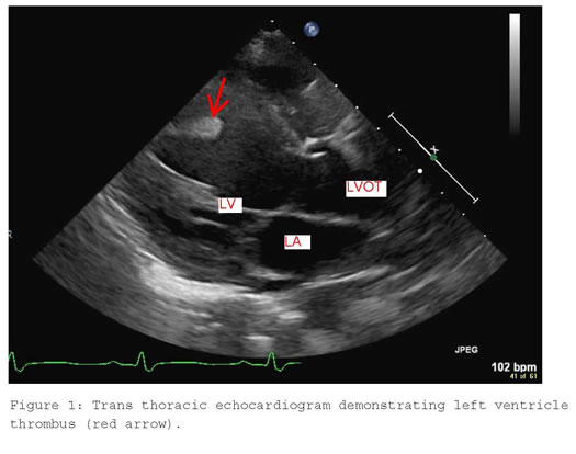
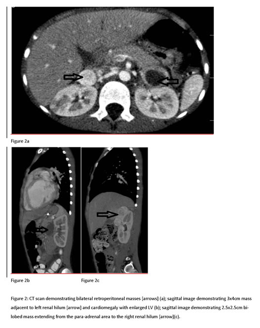
This is a previously healthy 9 year old boy who presented to his primary care provider with a two month history of recurring fevers, lethargy, and abdominal pain. The patient’s family medical history was significant for Von Hippel Lindau syndrome in his mother. A chest x-ray revealed cardiomegaly and pulmonary edema. An echocardiogram found a severely depressed ejection fraction of 27% as well as a large left ventricular thrombus. See Figure 1. An abdominal CT scan found bilateral retroperitoneal mass lesions, cystic masses in the abdomen, and evidence of emboli in the kidneys. See Figure 2. MRI of the brain demonstrated small foci of ischemic change consistent with embolic phenomenon.
The patient was admitted to an outside hospital, where he was started on intravenous infusions of milrinone and dopamine. Following clinical improvement, he was transitioned to an oral heart failure regimen. Despite the clinical improvement, a repeat echocardiogram demonstrated increased mobility in the left ventricular thrombus, mild mitral regurgitation, tricuspid regurgitation, and an ejection fraction of 17%. Subsequently the patient developed chest and arm pain accompanied by increasing troponins, ST-T wave changes and a new RBBB on electrocardiogram.
The patient was transferred to our facility, where he was placed on an infusion of milrinone for significantly diminished cardiac function. Given the patient’s family history and concerning CT scan, VMA and normetanephrine levels were sent to evaluate for pheochromocytoma. VMA was 44 mg/g (normal 0-9 mg/g). Normetanephrine was 4809 ug/24hr (normal 50-500 ug/24hr). Therapy with phenoxybenzomine, a long acting alpha receptor antagonist, was initiated. Carvedilol, a non-selective beta and alpha 1 blocker, was added, but the patient did not tolerate the orthostatic hypotension and bradycardia. A few days after his admission to the PCCU, the patient suffered a right MCA stroke resulting in left-sided weakness. The left ventricle thrombus was absent by ECHO exam. A multidisciplinary meeting was held to discuss options for managing the pheochromocytomas; the consensus was to pursue medical management, in light of the patient’s high perioperative risk with the hope that his cardiac function would improve.
Despite two months of medical management; consisting of phenoxybenzamine, furosemide, and milrinone, in an attempt to aggressively normalize blood pressure and decrease the amount of catecholamines, the patient’s cardiac function continued to decline. An echocardiogram demonstrated mild to moderate mitral regurgitation, severely depressed left ventricular wall motion, and mildly depressed right ventricular wall motion. The decision was made to proceed with surgical removal of two right adrenal tumors and one left adrenal tumor. Given the fragile cardiac function of the patient (now with an ejection fraction of 15%) it was unclear if the patient would tolerate induction of anesthesia and the hemodynamics swings associated with pheochromocytoma resection. The plan was to proceed with open surgical resection with invasive monitoring and to have ECMO on standby, including a second surgical team for cannulation, if hemodynamic instability ensued.
On the morning of surgery; the surgical, ECMO, and anesthesia teams were present in the OR prior to induction. Standard monitors, an existing left radial art line and cerebral oximetry monitors were placed. His blood pressure (BP) was 90-100/ 50-60 prior to and after induction. The patient was induced with etomidate, fentanyl and vecuronium. A central venous line, continuous cardiac output catheter and transesophageal echo were placed after induction.
Anesthesia was maintained with isoflurane, intermittent fentanyl and vecuronium.
The patient received a stress dose of steroids (50mg of hydrocortisone) shortly after induction. A milrinone infusion was maintained at 0.75 mcg/kg/min throughout the case. There was a rise in BP (up to 120s systolic and 70s diastolic) and HR (up to 160s) during manipulation of the right adrenal masses. Sodium nitroprusside (0.3-1 mcg/kg/min) was not effective in managing hypertension and tachycardia alone, so esmolol was added (50-150 mcg/kg/min). Ultimately, boluses of phentolamine (0.5mg), followed by an infusion at 0.1mg/kg/hr, was effective in managing increases in heart rate and blood pressure. After removal of the second adrenal mass, there was a decline in the BP to 70/50. E smolol, phentolamine, and nitroprusside were discontinued, and norephinephrine (0.1 – 0.2 mcg/kg/min) and vasopressin (0.6 - 2.5units/hr) were added for management of hypotension. The patient’s serum glucose dropped from a normal range of 110-120 to 50 following tumor excision requiring several boluses of D50 during the first post operative night.
The patient’s postoperative course was protracted and complicated by continued poor cardiac function. He was extubated on postoperative day 3 to CPAP. Milrinone was gradually weaned and discontinued on postoperative day 39. His echocardiogram completed prior to discharge was essentially unchanged from pre-op; with a severely dilated left ventricle with markedly depressed wall motion and moderate to severely dilated right ventricle with moderate to markedly depressed wall motion. He was discharged on post operative day 59 on an oral regimen of enalapril, digoxin, and carvedilol for his heart failure. He continues to require steroid replacement therapy.
Discussion
Classically, patients with pheochromocytomas present with episodic headaches, sweating and palpitations. Rarely, prolonged exposure of catecholamines directly acting on the myocardium may result in a dilated cardiomyopathy. Pheochromocytomas produce a myopathy that is indistinguishable from lesions induced in animal models given norephinephrine infusions suggesting that the resulting cardiomyopathy is mediated via an alpha-adrenergic pathway. Pretreatment with prazosin, an alpha 1 antagonist, reduces histological damage in rabbits infused with norepinephrine3. Imperato-McGinley et al. documented the reversibility of catecholamine-induced dilated cardiomyopathy with the use of MPT, a hydroxylase inhibitor that lowers plasma norephinephrine levels. However, they did note that MPT resulted only in moderate improvement of cardiac function. Dramatic improvement of cardiac function only occurred after surgical resection.
Once the diagnosis of pheochromocytoma induced cardiomyopathy was established, a multidisciplinary meeting to decide the course of therapy was held. The definitive therapy was felt to be surgical resection. However, there was concern for increased morbidity and mortality associated with pheochromocytoma resection in the presence of cardiomyopathy. The consensus of the group was to pursue medical therapy in the hope that his cardiac function would recover. Despite two months of aggressive inpatient medical therapy consisting of phenoxybenzamine for alpha blockade, a trial of carvedilol for beta blockade, and milrinone for inotropic support , our patient’s cardiac function continued to decline. Unfortunately, 2 months following resection, our patient’s cardiac function remained unchanged.
Cardiomyopathy is a rare, but not uncommon disorder in children. It has important anesthetic implications, requiring close attention to; maintenance of preload, maintenance of afterload reduction, avoidance of decreased myocardial contractility, and avoidance of elevated systemic vascular resistance. Adults with heart failure undergoing non-cardiac surgery have a twofold higher mortality and readmission rate than patients without heart failure. Less is known about anesthetic related and postoperative events in children with cardiomyopathy due to the paucity of literature5. Kipps et al. showed the need for escalated care pre- and post-procedure in children with severe ventricular dysfunction. Complications occurred in 83% of the children with severe ventricular dysfunction. Peri-procedural morbidity was noted in 38% of the procedures.
Here we describe the successful peri-operative management of the resection of bilateral pheochromocytomas in a pediatric patient with severe catecholamine induced cardiomyopathy refractory to medical therapy. The anesthesiologist must be mindful and aware of the complex physiological changes expected during pre and post surgical removal of the tumor. To prevent un-controlled intraoperative hemodynamics, pretreatment with an alpha antagonist such as phenoxybenzamine is essential. Invasive hemodynamic monitoring may aid in management of post tumor excision labiality, including hypotension and hypoglycemia.
References
- Eisenhofer, Graeme et al. Pheochromocytomas in von Hippel-Lindau Syndrome and Multiple Endocrine Neoplasia Type 2 Display Distinct Biochemical and Clinical Phenotypes
- Hack, H.A. The Perioperative Management of Children with Phaeochromocytoma. Paediatric Anaesthesia 2000; 10: 463-476.
- Imperato-McGinley, Julianne et al. Reversibility of Catecholamine-Induced Dilated Cardiomyopathy in a Child with a Pheochromocytoma. The New England Journal of Medicine 1987; 316: 793-797.
- Kinney, Michelle A.O. et al. Perioperative Management of Pheochromocytoma. Journal of Cardiothoracic and Vascular Anesthesia 2002; 16: 359-369
- Kipps, Alaina K. et al. Children with Cardiomyopathy: Complications After Noncardiac Procedures with General Anesthesia. Pediatric Anesthesia 2007; 17: 775-781.
- Matsota, P. et al. Anaesthesia for Phaeochromocytoma Removal in a 5-Year-Old Boy. Paediatric Anaesthesia 2002; 12: 176-180.
- McMillian, Wesley et al. Phentolamine Continuous Infusion in a Patient with Pheochromocytoma. American Journal of Health-System Pharmacy 2011; 68: 130-134
- Prys-Roberts, C. Phaeochromocytoma-Recent Progress in its Management. British Journal of Anaesthesia 2000; 85: 44-57
